
Radiology
Monroe Regional Hospital’s Department of Radiology offers state-of-the-art imaging in a professional and personal setting. Our radiology department is covered 24/7 by a licensed Radiology Technologist. Our studies are interpreted by a licensed Radiologist and most results will be relayed to your Physician within 24 hours. Monroe Regional Hospital is accredited by the American College of Radiology in CT and MRI. We are pleased to offer various radiology services including:
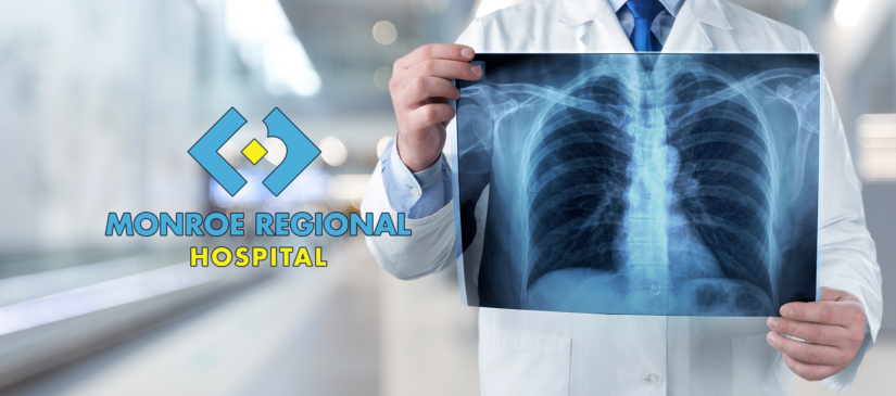
General Radiology (X-ray)
General radiology tests, also called x-rays, use high-energy radiation to create images of bones and internal organs. X-ray is a simple, painless medical test that helps your physician view and assess conditions ranging from broken bones to pneumonia to cancer.
Ultrasound
An ultrasound test uses reflected sound waves to produce an image of the organs in the body. The words “ultrasound study”, “ultrasound exam”, and “sonogram” all mean the same thing.
During an ultrasound the patient is asked to lie down on the exam table. A gel is applied to the part of the body being examined and then a small instrument called a transducer or probe is passed over that area by the sonographer. The patient may be asked to move into different positions or to hold their breath to improve imaging. Some ultrasound examinations require special preparations, which will be explained during scheduling.
CT (Computed Tomography or CAT Scan)
Computed Tomography is a type of radiologic exam where the x-ray source rotates around the patient. A computer then interprets the information and creates a cross-sectional image or a “slice”.
The patient is asked to lie on a table and the area being scanned will be moved into the scanner. For most exams the patient will lie on his or her back. Some of the exams require the patient to hold their breath. Also depending on the type of exam requested, an intravenous contrast may be administered. Preparation is required for some examinations, which may involve drinking an oral contrast and/or fasting. These preparations will be explained at the time of scheduling.
MRI
MRI is a way of taking pictures of your body and its chemical make-up. It’s a safe way to make an image of your body that does not use radiation. It uses a strong magnetic field supplied by a magnet and radio waves.
An MRI examination requires the patient to lie on a table. The area of the body to be scanned will be positioned in the center of the magnet. The patient will enter the magnet either feet or head first, depending on the type of exam being performed. A small device called a surface coil may be placed near the part of the body to be scanned to improve images.
Echocardiogram
An echocardiogram is an ultrasound test that can evaluate the structures of the heart, as well as the direction of blood flow within it. Technicians specially trained in echocardiography produce the images and videos, often using a special probe or transducer that is placed in various places on the chest wall, to view the heart from different directions. Cardiologists, or heart specialists, are trained to evaluate these images to assess heart function and provide a report of the results. The echocardiogram is just one of the many tests that can be done to evaluate heart anatomy and function.